< Page 12 of 15 >
1 2 3 4 5 6 7 8 9 10 11 12 13 14 15
Anatomical
Considerations
Staining Patterns
Corneal Epithelial Staining Patterns The loss of corneal epithelial cells is one of the most common adverse consequences of contact lens wear. Although this can be detected through the use of the slit lamp, the installation of flourescein dye can indicate the precise nature and location of specific corneal epithelial defects. Flourescein acts either by pooling in the area of the defect or by staining the underlying exposed basement membrane or Bowman�s layer. Corneal epithelial damage can result in the discrete loss of a few epithelial cells to deeper craterlike lesions with cell loss to the level of Bowman�s membrane. Epithelial damage can be caused either by direct trauma or by defective tear film distribution over the cornea.
Epithelial
Defects Induced by Direct Trauma:
-
A poorly edged or a damaged lens
-
An excessively flat lens
-
Foreign particles such as dust or cosmetics which lodge beneath the lens
-
Improper insertion, removal and recentering techniques
-
Poor cleaning habits
- Mucus buildup generally due to the dry storage of hard lenses
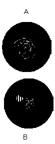
Figure A below illustrates a swirl-like stain, semicircular in configuration. Possible causes are keratoconus associated with apical touch, a flat lens-cornea relationship, or a poorly centered lens. Figure B illustrates foreign body stains which can leave either a thin snakelike track on the cornea as it travels under the lens or a small crater-like lesion as the result of becoming embedded in the cornea. The most common foreign matter to be trapped under the lens is dust, followed by eye makeup and mucus particles.
< Page 12 of 15 >
1 2 3 4 5 6 7 8 9 10 11 12 13 14 15