< Page 3 of 15 >
1 2 3 4 5 6 7 8 9 10 11 12 13 14 15
Anatomy Review
Eyelids:
The eyelids
function primarily to help keep the eye moist and to help keep out foreign
bodies. It is the opposing action of the eyelids which causes tears
to spread evenly over the cornea thus keeping it moist. The lids play
an important role in the fitting and wear of contact lenses for the
following reasons: They effect the wetability of the lens surface They
effect the positioning of the lens They are sensitive and generally
cause most of the discomfort when a lens is first placed on the eye
Conjunctiva:
The conjunctiva
is the loose tissue covering the sclera and inside the lids. The bulbar
conjunctiva covers the sclera while the palpebral conjunctiva is that
portion which lines the inner surface of the upper and lower eyelids.
Cornea:
The cornea
is the anterior refracting surface of the eye, it consists of transparent
tissue and is devoid of blood vessels. The cornea is of utmost importance
to the contact lens fitter since the lens sits directly on this tissue.
The average corneal thickness is about 0.52 mm at its center and increases
to about 0.65 mm at the limbal area and it is composed of five distinct
layers.
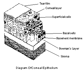
Epithelium: That layer of the cornea which is exposed to the tears and comprises about 10 % of the total corneal thickness. The corneal epithelium is highly regenerative, demonstrating a remarkable ability to heal itself after being scratched. A corneal abrasion is often completely healed within 24 hours.
Bowman�s Membrane: Is essentially a modification of the underlying stroma. Unlike the epithelium, if damaged by a scratch or cut, it can not regenerate itself so scarring will occur. s
Stroma:The stroma comprises approximately 90% of the corneal thickness. It consists of 200 to 250 layers of cells called lamellae, which lie parallel to the corneal surface. If damaged by injury, scarring will occur which can result in opacities of the cornea. Chronic infection or corneal edema can cause blood vessels to invade the stroma resulting in a condition known as neovascularization. These blood vessels which enter to supply oxygen and nutrients can obscure vision.
Descemet�s Membrane: A strong structureless layer which is secreted by the endothelium. It is elastic, and resistant to trauma and pathology.
Endothelium: The innermost or most posterior layer of the cornea, consisting of a single layer of flattened cells. In contrast toDescemet�s Membrane, these cells are very susceptible to trauma and pathology. And in marked contrast to the epithelium, endothelium cells are infrequently, if ever replaced as a normal process during adult life. If disrupted, however, they can be replaced by the spreading of healthy cells.
Limbus:
The limbus
is a transition zone between the cornea and the sclera. It is approximately
1 mm wide and the cornea is dependent upon it for receiving part of
its nutrients. The limbal region becomes significant when fitting contact
lenses since it is so closely connected to the cornea and some contact
lenses will bear directly on it.

< Page 3 of 15 >
1 2 3 4 5 6 7 8 9 10 11 12 13 14 15